
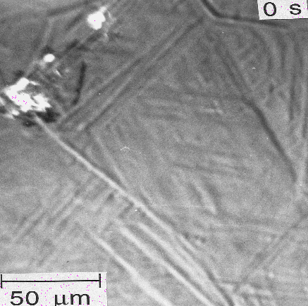
The lengthening rate of individual bainite platelets has been measured using hot-stage photoemission electron microscopy. Electrons are excited from the surface of the sample using incident ultraviolet radiation, and it is these photo-emitted electrons which form the image. The technique can resolve individual platelets of bainite. The movie shown below illustrates a series of photoemission electron micrographs taken at 1 s intervals, showing the growth of the plates. The measured lengthening rate of the arrowed sub-unit is 75 microns per second. This is many orders of magnitude greater than expected if growth is assumed to occur by a paraequilibrium carbon diffusion-controlled rate (0.083 microns per second). Lengthening therefore occurs much faster than expected from carbon diffusion-control growth.
For details see Kinetics, Solute-Drag and Mechanism of the Bainite Reaction in Steels, by H. K. D. H. Bhadeshia, Proceedings of an International Conference on Phase Transformations in Ferrous Alloys, eds A. Marder and J. Goldstein, Philadelphia, A.I.M.E., 1984, pp. 335-340. It is a pleasure to acknowledge the help of Marimuthu Murugananth for the production of this movie from the images presented in the paper.
| PT Group Home | Materials Algorithms |

|

|